Description
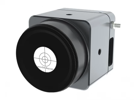
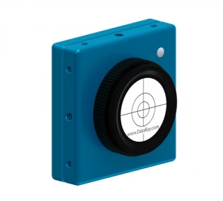
With a large 25 x 25 mm active area, 4.2 Mpixels, 12.5 x 12.5 μm (effective) pixels, optical and electronic triggering of a global shutter, and an SNR of 2500:1, the TaperCamD-LCM laser beam profiler offers the largest active sensor area on a USB-port powered laser beam profiling device. By combining the high signal-to-noise ratio and global shutter of the WinCamD-LCM with a high-quality fiber optic taper, the TaperCamD-LCM beam profiler offers a very compact, easy-to-use solution for measuring a variety of large CW or pulsed lasers.
The TaperCamD-LCM beam profiler is paired with DataRay’s full-featured, highly customizable, user-centric software (which has no license fees, unlimited installations, and free software updates). It is perfect for applications including: CW and pulsed laser profiling; field servicing of laser systems; optical assembly; instrument alignment; beam wander and logging; R&D; OEM integration; and quality control.
TaperCamD-LCM CMOS Based Beam Profiler
Specifications
| Sensor Type: | CMOS |
|---|---|
| Measurable Sources: | CW, Pulsed |
| Wavelength Range: | 355 – 1150 nm |
| # Pixels (Width): | 2048 |
| # Pixels (Height): | 2048 |
| Pixel Size (Width): | 12.5 um |
| Pixel Size (Height): | 12.5 um |
| Max Full Frame Rate: | 60 Hz |
| ADC: | 12-bit |
| Part Number: | S-TCD-LCM |
| Beam Fits: | Gaussian & Top Hat profile fit & % fit Equivalent Slit profile |
| Centroid Position: | Relative and absolute | Intensity Weighted Centroid and Geometric Center | Beam Wander Display and Statistics |
| Measured & Displayed Profile Parameters: | Raw and smoothed profiles Triangular running average filter up to 10% FWHM |
| Displayed Profiles: | Line‚ 2D & 3D plots. Normalized or un-normalized. Linear or Logarithmic‚ Zoom x10 2D‚ 3D in 10‚ 32 or max. colors or grayscale Contoured display at 10 and 32 colors |
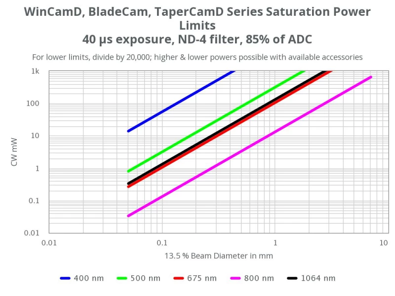
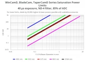
| Manual Beam Attenuation: | Includes three 2" NDXL filters (NDXL-1, NDXL-2, NDXL-4) |
| Measurable Sources: | CW beams‚ pulsed sources; CW to 12.6 kHz with single pulse isolation Software configurable Auto-trigger‚ Synchronous & Variable Delay |
| ADC: | 12-bit |
| Electronic Shutter Range: | USB 2.0: 12,600:1 (41 dB) | USB 3.0: 25,000:1 (44 dB) |
| Signal To RMS Noise: | 2,500:1 (34 dB optical / 68 dB electrical) |
| Single Pulse Capture PRR: | USB 2.0: 6.3 kHz | USB 3.0: 12.6 kHz |
| Shutter Type: | Global |
| Min. Beam (10 Pixels): | ~125 µm |
| Image Area: | 25 x 25 mm |
| Measurement Accuracy (not Limited To Pixel Size): | 0.1 µm processing resolution for interpolated diameters. | Absolute accuracy is beam profile dependent - ~1 µm accuracy is frequently achievable. | Centroid accuracy is also beam dependent (as good as ±1 µ since it is arithmetically determined from all pi |
| Processing Options: | Image & profile Averaging‚ 1‚ 5‚ 10‚ 20‚ Continuous. | Background Capture and Subtraction. | User set rectangular Capture Block for capture | User set or Auto ellipse Inclusion region with beam tracking for processing | *.ojf files save all WinCamD custom |
| Pass/Fail Display: | On-screen selectable Pass/Fail colors. Ideal for QA & Production. |
| Log Data And Statistics: | Min.‚ Max.‚ Mean‚ Standard Deviation‚ to 4096 samples |
| Relative Power Measurement: | Rolling histogram based on user's initial input. Units of mW‚ µJ‚ dBm‚ % or user choice (relative to a reference measurement input) |
| Fluence: | Fluence‚ within user defined area |
| Certification: | RoHS‚ WEEE‚ CE |
| Multiple Cameras: | Up to 4 cameras, parallel capture. | 1 to 8 cameras, serial capture. |
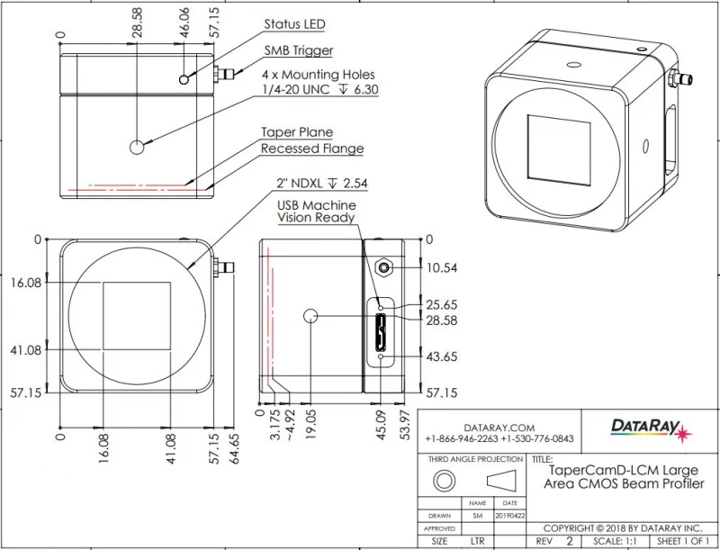
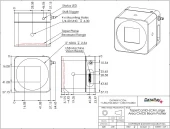
| Head Dimensions‚ Width X Height X Depth: | 2.25 x 2.25 x 2.13” (57 x 57 x 54 mm) |
| Mounting: | 1/4-20 thread |
| Weight‚ Camera W/ NDXL And Filter Cover: | 15.2 oz (431 g) |
| Beam Ellipticity: | Major‚ Minor & Mean diameters. Auto-orientation of axes. |
| Beam Diameter: | Diameter at two user set Clip levels Gaussian & ISO 11146 Second Moment beam diameters Equivalent diameter above a user defined Clip level Equivalent Slit and Knife Edge diameters |
Got questions about specs? Use the inquiry form to ask.
Features
- 355 to 1150 nm, standard CMOS detector
- 4.2Mpixel, 2048 x 2048 pixels, 25 x 25 mm active area
- 12.5 µm pixels (effective)
- HyperCal™ – Dynamic noise and baseline correction software
- Port-powered USB 3.0; flexible screw locking 3 m cable; no power brick
- 12-bit ADC, on-board microprocessor
- Window-free sensors standard for no fringing
- 25,000:1 electronic auto-shutter, 79 µs to 2 s
- 2,500:1 SNR
- Global shutter, optical/TTL trigger
- Isolated pulse triggering and parallel capture
Note: This product requires 64-bit Windows 7, 8/8.1, or 10
Applications
- CW and pulsed laser profiling
- Field servicing of lasers and laser-based systems
- Optical assembly & instrument alignment
- Beam wander & logging
- Alignment and diagnostics of laser systems used in ophthalmology, dermatology, or endoscopic surgery
- Used experiments involving SHG, OPOs, or other nonlinear processes that require beam control at tight foci
- Useful in development and QA of optical read/write heads where small, highly focused beams are critical
- Enables spatial profiling of tightly focused beams from laser diodes and VCSELs used in sensing or communication
- Assists in beam shaping and optimization in advanced microscopy systems for biomedical imaging and dimensional metrology
Frequently Asked Questions
What are the applications of the TaperCamD-LCM beam profiler?
The TaperCamD-LCM beam profiler is perfect for applications including CW and pulsed laser profiling, field servicing of laser systems, optical assembly, instrument alignment, beam wander and logging, R&D, OEM integration, and quality control.
What is the active area of the TaperCamD-LCM beam profiler?
The TaperCamD-LCM beam profiler has a large 25 x 25 mm active area.
What is the frame rate of the TaperCamD-LCM beam profiler?
The TaperCamD-LCM beam profiler has a frame rate of 60 fps @ 512 x 512, 30 fps @ 1024 x 1024, 12 fps @ 2048 x 2048.
What is the required operating system for the TaperCamD-LCM beam profiler?
The TaperCamD-LCM beam profiler requires 64-bit Windows 7, 8/8.1, or 10.
What is the SNR of the TaperCamD-LCM beam profiler?
The TaperCamD-LCM beam profiler has an SNR of 2500:1.
Does the TaperCamD-LCM support external triggering for synchronized measurements?
Yes, the TaperCamD-LCM features a global shutter with both optical and TTL trigger inputs, allowing for precise synchronization with external events or pulsed laser sources.
What is the minimum beam size that the TaperCamD-LCM can accurately profile?
The TaperCamD-LCM can accurately profile beams with diameters as small as approximately 125 µm, ensuring detailed analysis of small beam spots.
Is the TaperCamD-LCM compatible with different operating systems?
The TaperCamD-LCM is compatible with 64-bit versions of Windows 7, 8/8.1, and 10, providing flexibility for integration into various system environments.
Got more questions? Use the RFQ form to ask the supplier directly.
Similar Products
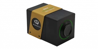
BeamPro | Compact Footprint Laser Beam Profiler
Axiom Optics
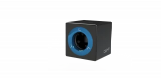
BeamPro | Large Area Laser Beam Profiler
Axiom Optics
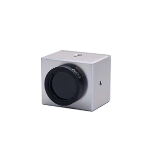
BeamPro | Small Pixel Laser Beam Profiler
Axiom Optics
Wavefront Sensor Based Laser Beam Profiler - SID4
PHASICS
CaM² M-Squared meter
Imagine Optic
CMOS Beam Profiler
YIXIST YIXI Technology Co., Ltd
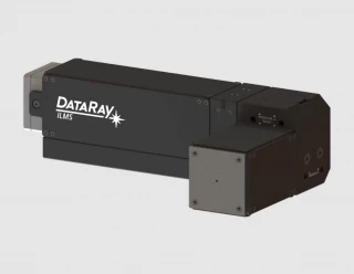
Industrial Laser Monitoring System (ILMS)
DataRay Inc.

BladeCam2-XHR – ½" CMOS Beam Profiler System
DataRay Inc.
BladeCam2-HR – ½" CMOS Beam Profiler System
DataRay Inc.
WinCamD-QD – Quantum Dot SWIR Beam Profiler
DataRay Inc.
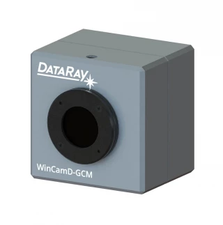
WinCamD-GCM 1" - GigE Vision CMOS Beam Profiling Camera
DataRay Inc.
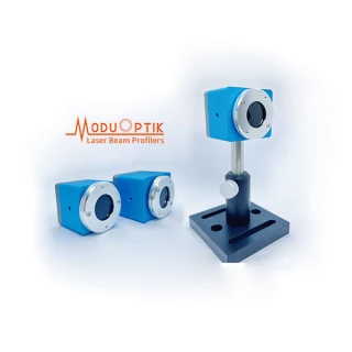
ModuOptik - Beam Profiler (CMOS)
Hefei ModuOptik Technologies Co., Ltd.
Need pricing for this product? Send a quick inquiry
Thank You!
Your inquiry has been received.
Create an account by adding a password
Why create an account?
- Auto-complete inquiry forms
- View and manage all your past messages
- Save products to your favorites
- Close your account anytime — no hassle