
Description
LUXYR LED PICO – Compact Direct-Fit LED Illumination for Demanding Microscopy
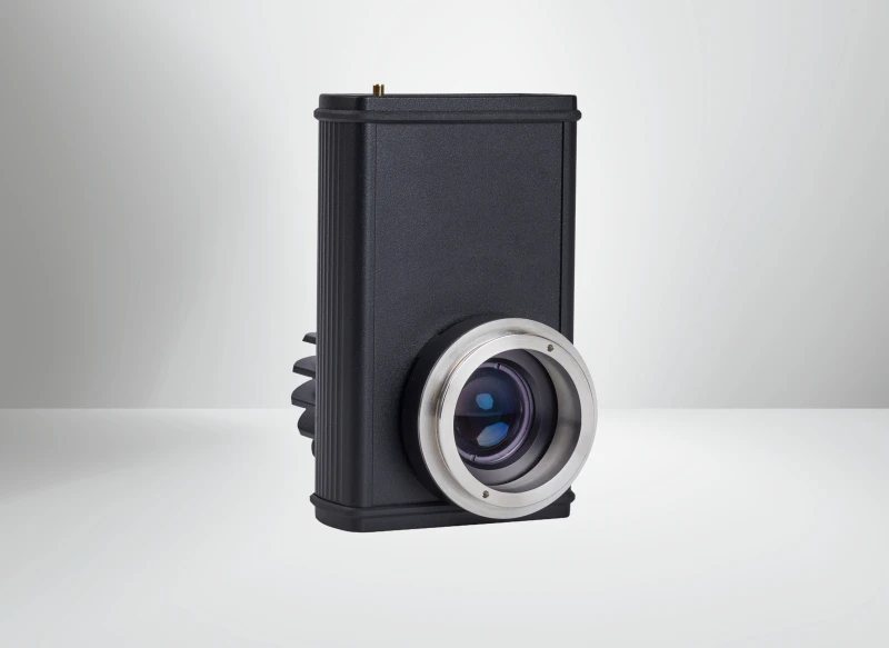
The LUXYR LED PICO is a high‑performance, direct-fit LED light source engineered for modern fluorescence and brightfield microscopy. Designed as a compact replacement for conventional halogen or mercury lamps, it combines high radiant output with excellent illumination homogeneity in a form factor that integrates directly into the microscope’s illumination path. With versions spanning UV‑A to near‑infrared (365–735 nm) as well as high‑intensity white‑light models, this system provides a flexible platform for laboratories and OEMs looking to precisely match their light source to specific imaging tasks.
In contrast to traditional arc lamps, the LED PICO delivers instant-on operation, long service life, and stable output without warm‑up times or bulb replacements. Its collimation optics and dedicated microscope adapters are optimized for coupling efficiency and uniform field illumination across the specimen, making it suitable for both routine imaging and advanced quantitative applications. For users upgrading older microscopes, the direct‑fit design offers a straightforward path to LED illumination with minimal mechanical changes and no compromise in optical performance.
Broad Spectral Range from UV-A to NIR and High-Quality White Light
The LED PICO is available in monochromatic variants from 365 nm to 735 nm, covering UV‑A, visible, and NIR regions important for a wide variety of fluorophores and inspection tasks. Typical lines include 365 nm, 385 nm, 405 nm and other application-relevant wavelengths, each chosen to provide strong radiant power and narrow spectral output for efficient fluorophore excitation and reduced background. These discrete wavelengths make it easy to optimize contrast and signal‑to‑noise ratios in fluorescence microscopy, darkfield imaging, and specialized inspection workflows.
In addition to monochromatic options, the LED PICO is available as a powerful white‑light source with integral luminous flux up to 2500 mW and luminous output up to 800 lm. HighFlux White versions provide intense illumination for demanding brightfield and darkfield microscopy, while HighCRI White variants (up to CRI 90) are tailored for applications requiring accurate color rendering, such as pathology, life‑science imaging, and visual inspection. This combination of output power and spectral quality makes the LED PICO competitive with or superior to many conventional microscope illuminators in both research and industrial settings.
Precise Electronic Control for Continuous, Follow, and Pulsed Operation
A key strength of the LED PICO is its sophisticated electronic control, designed to support both stand‑alone use and tight integration into automated imaging systems. In continuous‑wave (CW) mode, the device operates with adjustable luminous flux and minimal light ripple (< 0.5 %), delivering stable, flicker‑free illumination for visual observation, long‑exposure imaging, and quantitative measurements. Brightness control is gamma‑corrected, so perceived intensity changes are smooth and linear from the user’s perspective, which is particularly helpful when reproducibility and fine intensity tuning are required.
For synchronized imaging or high‑speed acquisition, the LED PICO supports advanced triggering modes via a TTL interface. In Follow mode, the light output is level‑controlled by a TTL signal, offering fast rise and fall times (t10‑90 and t90‑10 < 2 µs) and signal runtimes under 1 µs, enabling precise coordination with camera exposure or scanning systems. In Pulse mode, edge‑controlled pulses can be configured with defined delay, pulse length, and luminous flux. Minimum pulse length is on the order of 10 µs with delays below 5 µs, making this light source suitable for time‑critical applications such as high‑speed fluorescence imaging, stroboscopic inspection, and triggered machine‑vision setups.
Flexible Integration and Silent Operation for Sensitive Environments
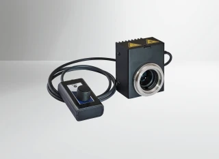
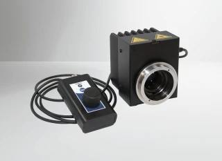
The LED PICO has been designed with integration flexibility in mind. It features a compact housing (approximately 134 mm × 85 mm × 97 mm) and a low weight of around 0.8 kg, making it easy to mount on a broad range of upright and inverted microscopes or to incorporate into custom optical systems. Interchangeable microscope adapters are available for major microscope brands such as Zeiss, Leica, Nikon, and Olympus, with the option for customized adapters to fit OEM or specialty instruments. Integrated collimation optics ensure that the light is efficiently directed through the microscope’s illumination path, reducing setup time and alignment effort.
LUXYR LED PICO Microscope Light Source UV to NIR and White Illumination
Specifications
| Light Source Technology: | LED |
|---|---|
| Spectral Output: | White / broadband, Monochromatic, Multi-wavelength |
| Wavelength Range: | 365 – 735 nm |
| Number Of Channels: | 1 |
| Microscope Coupling: | Direct-fit adapter |
| Compatible Microscopes: | Zeiss, Leica, Nikon, Olympus/Evident, Custom |
| Application: | Fluorescence, Brightfield, Darkfield, Forensics, Inspection / machine vision, Metrology |
| External Control: | TTL, Control panel only |
| Fall Time (pulse Mode): | < 2 µs (t90–10) |
| Accessories: | Configuration software |
| Scope Of Delivery: | Light source, control panel, power supply, microscope adapter of choice, operating instructions |
| Weight: | 0.8 kg |
| Dimensions (light Source): | 134 mm × 85 mm × 97 mm (L/H/W) |
| Power Consumption: | max. 36 VA |
| Mains Voltage (power Supply Unit): | 100–240 VAC ± 10%, 50–60 Hz |
| Optical Output: | Collimation optics with microscope adapter (Zeiss, Leica, Nikon, Olympus, customized) |
| DC Input: | Hollow plug (power supply unit) |
| TTL Interface: | SMB socket (follow & pulse mode) |
| USB Interface: | USB 2.0 type B micro (configuration, operation, status) |
| Control Interface: | Mini-DIN (control panel) |
| Minimum Delay: | < 5 µs |
| Minimum Pulse Length: | 10 µs (tPulse min) |
| Light Source Type: | Direct-fit LED, monochromatic 365–735 nm or white-light |
| Rise Time (pulse Mode): | < 2 µs (t10–90) |
| Signal Runtime (follow Mode): | < 1 µs |
| Fall Time (follow Mode): | < 2 µs (t90–10) |
| Rise Time (follow Mode): | < 2 µs (t10–90) |
| TTL Trigger Frequency: | up to 100 kHz |
| Light Ripple (CW Mode): | < 0.5% |
| Peak Wavelength 405: | 405 nm ± 5 nm, > 1700 mW |
| Peak Wavelength 385: | 385 nm ± 5 nm, > 1700 mW |
| Peak Wavelength 365: | 365 nm ± 5 nm, > 1200 mW |
| CRI (HighCRI White): | CRI 90 |
| CRI (HighFlux White): | CRI 70 |
| Integral Luminous Flux (HighCRI White): | > 2500 mW (700 lm) |
| Integral Luminous Flux (HighFlux White): | > 2500 mW (800 lm) |
Got questions about specs? Use the inquiry form to ask.
 Download Data Sheet

Features
- Direct-Fit LED Light Source for Microscopy – Compact, plug-in replacement for conventional mercury and halogen light sources in fluorescence and brightfield microscopes, reducing system complexity and maintenance.
- Wide Spectral Range: UV-A to NIR + White – Monochromatic LED options from 365 nm to 735 nm as well as high-flux and high-CRI white versions, enabling optimal excitation for a broad range of fluorophores and contrast techniques.
- High Optical Output for Demanding Imaging – Up to 2,500 mW radiant power or 800 lm luminous flux (white version), suitable for high-sensitivity fluorescence, fast camera-based imaging, and low-noise brightfield imaging.
- Homogeneous, Stable Illumination – Integrated collimation optics provide uniform field illumination across the microscope’s field of view, improving quantitative image analysis and reproducibility.
- Gamma-Corrected Brightness Control – Precise, linear-feeling intensity adjustment across the entire dimming range allows accurate setting of exposure levels for both visual observation and camera-based imaging.
- Advanced Operating Modes (CW, Follow & Pulse) – Continuous-wave mode for steady illumination, level-controlled “follow mode” (TTL) with predefined intensity, and edge-triggered pulse mode with configurable delay, pulse length and luminous flux.
- High-Speed TTL Triggering up to 100 kHz – TTL input via SMB socket allows rapid on/off switching and stroboscopic illumination, ideal for time-resolved fluorescence, high-speed imaging and synchronized machine-vision applications.
- Fast Rise and Fall Times (< 2 µs) – Very short turn-on/turn-off times (t10–90 < 2 µs, t90–10 < 2 µs) enable precise temporal control, reducing photobleaching and phototoxicity by limiting sample exposure to the actual acquisition time.
- Very Short Minimum Pulse Length (10 µs) – Edge-controlled pulse mode supports minimum pulse lengths of 10 µs and delays < 5 µs, providing excellent flexibility for synchronized imaging sequences.
- Low-Ripple Output for Image Stability – Continuous operation with light ripple < 0.5% ensures flicker-free, stable illumination, which is crucial for quantitative fluorescence measurements and high-dynamic-range imaging.
- Silent, Vibration-Free Operation – Fanless (or ultra-quiet) solid-state design operates without noticeable noise or mechanical vibration, protecting delicate setups (e.g., live-cell microscopy, long-exposure imaging).
- Compact Footprint & Lightweight Housing – Small form factor of approx. 134 mm × 85 mm × 97 mm (L × H × W) and weight of about 0.8 kg saves valuable bench space and simplifies integration into OEM instruments and retrofit upgrades.
- Interchangeable Microscope Adapters – Compatible with leading microscope brands (Zeiss, Leica, Nikon, Olympus) and customizable adapters, making it easy to adapt the light source to existing research and industrial microscope platforms.
- Flexible Control Interfaces – Control via dedicated mini‑DIN control panel, USB 2.0 (Micro‑B) for configuration, operation and status feedback, plus TTL input for hardware triggering, allowing seamless integration into modern imaging workflows.
- USB Configuration & OEM-Friendly Software Tools – Configuration software (accessory) via USB enables custom settings (intensity, pulse parameters, operating modes) and supports integration into automated microscopy and inspection systems.
- Multiple LED Versions for Application-Specific Optimization – Choices include high-flux white (CRI ~70, up to 800 lm), high-CRI white (CRI ~90, ~700 lm) and monochrome 365, 385, 405 nm and other wavelengths on request, matching specific fluorophores and contrast methods.
- Energy-Efficient Solid-State Illumination – Maximum power consumption of approx. 36 VA with long LED lifetime significantly reduces operating costs and frequent lamp replacements typical with mercury or halogen sources.
- Broad Application Versatility – Ideal for fluorescence, brightfield and darkfield microscopy, as well as image processing, optical inspection, semiconductor manufacturing, quality control, and forensic microscopy.
- Global Mains Compatibility – External power supply operates from 100–240 VAC, 50–60 Hz, making the system suitable for worldwide deployment in research institutes, OEM instruments and industrial installations.
Applications
- Fluorescence Microscopy in Life Sciences – Optimized UV-A to visible LED options (365–405 nm and beyond) provide high excitation efficiency for common fluorophores (e.g., DAPI, FITC, TRITC, Cy5), enabling sensitive fluorescence imaging in cell biology, histology, immunofluorescence, and live-cell research.
- Brightfield Microscopy for Clinical and Research Labs – High-flux white LED versions (up to 800 lm) deliver homogeneous, stable Kohler-like illumination for routine pathology, hematology, cytology, and microbiology, replacing conventional halogen light sources in upright and inverted microscopes.
- Darkfield and Phase Contrast Microscopy – Uniform, flicker-free illumination improves contrast for unstained, transparent specimens such as live cells, microorganisms, and fine structures in materials, making subtle detail easier to detect in darkfield and phase contrast modalities.
- Fluorescence Stereomicroscopy and Macroscopy – Compact form factor and interchangeable microscope adapters (Zeiss, Leica, Nikon, Olympus, custom) support stereo microscopes for imaging larger samples such as embryos, small animals, insects, plant tissues, and fluorescently tagged model organisms.
- Epifluorescence Upgrades for Legacy Microscopes – Direct-fit LED design allows simple retrofitting of older halogen or mercury-based epifluorescence microscopes with modern solid-state illumination, increasing reliability, reducing maintenance, and eliminating hazardous lamps.
- Multicolor Fluorescent Imaging and Channel Switching – Availability of different monochromatic LEDs (365–735 nm) supports multi-channel imaging setups (UV, blue, green, red, NIR) for colocalization studies, FISH, GFP/RFP imaging, and multiplex labeling in biomedical and neuroscience research.
- Live-Cell and Time-Lapse Imaging – Stable, vibration-free, and low-heat LED operation is ideal for long-term experiments in incubated environments, minimizing thermal drift and phototoxicity while enabling precise timing via TTL triggering up to 100 kHz.
- Optogenetics and Photoactivation Experiments – Fast rise/fall times (< 2 µs) and TTL pulse control enable accurate light stimulation protocols for optogenetic actuators and photoactivatable fluorophores, supporting neuroscience, cardiac research, and functional imaging.
- High-Speed Fluorescence Imaging – Gamma-corrected brightness control and short minimum pulse length (10 µs) support synchronization with high-speed cameras and scanners in fast dynamic imaging, flow-based assays, or rapid tracking experiments.
- Forensic Microscopy and Crime Scene Analysis – UV and visible monochromatic LEDs (365, 385, 405 nm and others) enhance contrast and fluorescence of latent fingerprints, bodily fluids, fibers, and document forgeries under forensic microscopes and inspection stations.
- Quality Control in Semiconductor Manufacturing – Highly stable, homogeneous illumination supports inspection of wafers, photomasks, lithography features, bonded wires, and microstructures in brightfield, darkfield, and fluorescence-based defect detection.
- Electronic Component and PCB Inspection – Directionally stable LED output improves visualization of solder joints, microcracks, conformal coatings, and component placement under inspection microscopes used in electronics manufacturing and repair.
- Machine Vision and Automated Optical Inspection (AOI) – Precise control via USB and TTL with high repetition rates supports integration into automated imaging systems for part verification, pattern recognition, dimensional measurement, and surface defect detection.
- Industrial Metrology and Surface Analysis – Uniform white and monochromatic LED illumination is suitable for 2D and 3D surface measurements, roughness analysis, scratch and defect localization on precision mechanical parts, glass, and optical components.
- Material Science and Metallography – Brightfield and darkfield illumination support analysis of grain boundaries, phases, inclusions, and cracks in metals, ceramics, composites, and polymers, enabling reproducible imaging for research and quality control.
- Geology, Mineralogy, and Petrography – Stable white-light illumination assists in thin-section microscopy of rocks and minerals. UV/visible LEDs can be used to highlight fluorescent minerals and inclusions for identification and classification.
Frequently Asked Questions
What is the LUXYR LED PICO light source and what applications is it designed for?
Which LED versions and wavelengths are available for the LED PICO, and how do I choose the right one?
How is the LED PICO controlled and what operating modes are available (CW, Follow, Pulse)?
How do I integrate the LED PICO with my microscope or imaging system?
What are the power, size, and environmental characteristics of the LED PICO?
What is included in the scope of delivery and what software or accessories are available?
How does the LED PICO compare to traditional halogen or mercury lamp illumination in microscopy?
Got more questions? Use the inquiry form to ask the supplier directly.
Similar Products
Your inquiry has been received.
Create an account by adding a password
Why create an account?
- Auto-complete inquiry forms
- View and manage all your past messages
- Save products to your favorites
- Close your account anytime — no hassle