
Description
LED NANO High-Performance VIS LED Light Source for Microscopy
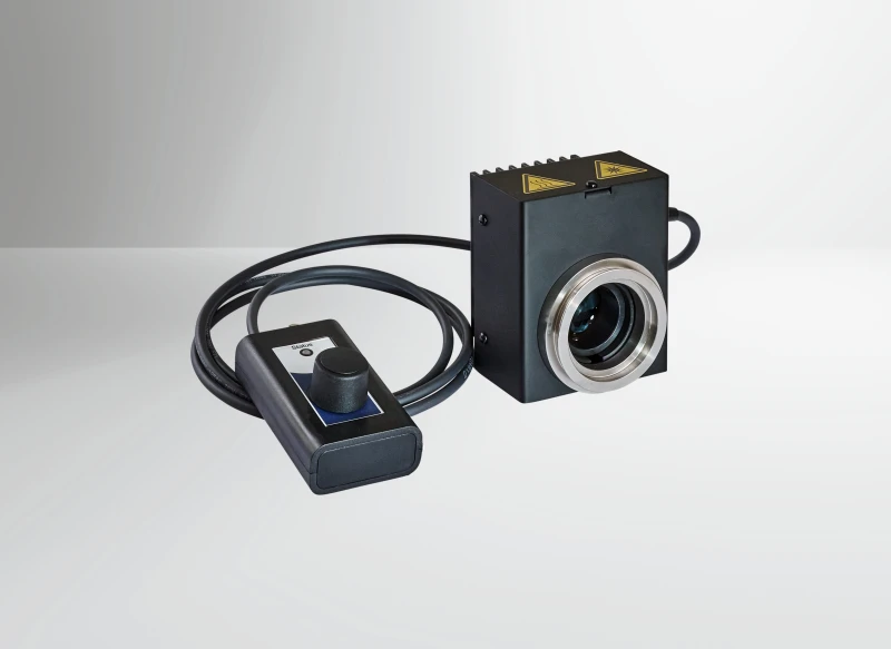
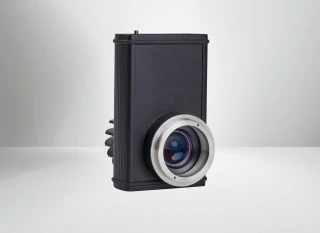
The LUXYR LED NANO is a highly compact, high-performance visible (VIS) LED light source engineered as a direct-fit replacement for traditional halogen illuminators in microscopy. Designed for the 400–780 nm spectral range, it delivers bright, homogenous illumination suitable for a wide array of optical techniques while dramatically reducing heat, noise, and maintenance compared with conventional lamps. With its small footprint and integrated collimation optics, the LED NANO fits seamlessly into modern and legacy microscope systems, offering a future-proof solution for research, industrial, and routine microscopy.
Unlike halogen light sources that suffer from short lamp lifetimes, significant heat output, and fluctuating intensity, the LED NANO offers a long LED lifetime of more than 20,000 hours and highly stable output. Users can choose between versions with a color rendering index (CRI) of 80 or 90, providing integral luminous flux up to approximately 350 lm. This allows optimization either for maximum brightness (CRI80) or superior color fidelity (CRI90), making it well suited for applications where color-critical assessment and reproducible image documentation are essential. The color temperatures around 3000 K and 4000 K are chosen to replicate or improve upon the visual impression of traditional halogen light, easing the transition to LED illumination.
The LED NANO has been purpose-built as a compact VIS light engine for brightfield, darkfield, forensic, material, and multispectral microscopy. Comparable to other advanced LED microscopy light sources found in modern imaging labs, it provides stable, broad-spectrum white light that effectively covers the visible range without the need for frequent bulb changes or warm-up times. Its optical design, including collimation optics and matched microscope adapters, ensures efficient coupling into the microscope, thereby maximizing light output at the sample while maintaining even illumination across the field of view. This results in improved imaging consistency for visual inspection, digital imaging, and automated analysis routines.
A key advantage of the LED NANO is its silent, vibration-free operation. In many microscopy setups—particularly in high-magnification, long-exposure, or low-light imaging—mechanical vibrations and fan noise from traditional light sources can degrade image quality and interfere with sensitive measurements. By employing a fanless design and efficient thermal management, the LED NANO maintains stable operating conditions without introducing mechanical disturbances. This contributes to sharper images, higher repeatability, and a more comfortable working environment, especially in laboratories that operate microscopes for extended periods.
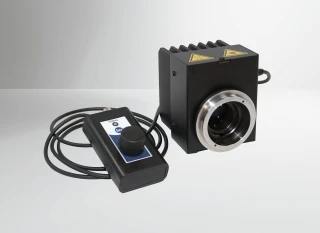
The device incorporates both local and external control options to integrate smoothly into existing workflows. A permanently attached control panel allows straightforward, manual adjustment of luminous flux in continuous (CW) operation. Thanks to low ripple of less than 0.5%, the light output remains extremely stable, which is particularly beneficial for quantitative imaging, documentation, and comparative analysis across multiple sessions. For automated or synchronized imaging setups, the LED NANO can also be externally switched via a TTL signal through a BNC interface. In follow mode, it offers level-controlled operation with preconfigured luminous flux and very short rise and fall times in the microsecond range, enabling precise timing with cameras, shutters, or scanning systems.
From a system integration perspective, the LED NANO has been developed with flexibility in mind. It supports interchangeable microscope adapters tailored for leading microscope brands such as Zeiss, Leica, Nikon, and Olympus, as well as customized adapters for OEM and specialized configurations. This modular approach allows the same light source to be used across different microscope families or to be deployed in dedicated imaging instruments. Compact dimensions of approximately 77 mm × 93 mm × 74 mm and a weight of about 0.5 kg including the control panel facilitate easy installation in crowded lab benches, integrated workstations, or instrument housings.
The power requirements (12 V DC, max. 18 VA) and the streamlined supply connector simplify integration into both standalone laboratory environments and OEM instruments. When compared to other LED-based microscopy illuminators used worldwide, the LED NANO stands out for its combination of small form factor, quiet operation, and flexible control options. Its efficient VIS LED technology helps reduce operating costs and downtime while delivering consistent illumination for demanding imaging tasks in biology, materials science, forensics, quality control, and multispectral imaging workflows.
Luxyr LED Nano Compact VIS LED Light Source for Microscopy Halogen Replacement
Specifications
| Light Source Technology: | LED |
|---|---|
| Spectral Output: | White / broadband |
| Wavelength Range: | 400 – 780 nm |
| Number Of Channels: | 1 |
| Microscope Coupling: | Direct-fit adapter |
| Compatible Microscopes: | Zeiss, Leica, Nikon, Olympus/Evident, Custom |
| Application: | Fluorescence, Brightfield, Darkfield, Forensics, Inspection / machine vision |
| External Control: | TTL, Control panel only |
| Operating Mode (Follow): | Level-controlled operation (TTL) with preconfigured luminous flux |
| Weight: | Approx. 0.5 kg (incl. control panel) |
| Dimensions (W×H×L): | 77 mm × 93 mm × 74 mm |
| Power Consumption: | Max. 18 VA |
| Voltage Supply: | 12 V DC |
| Supported Microscopes: | Zeiss, Leica, Nikon, Olympus, customised |
| Optical Output: | Collimation optics with microscope adapter |
| Supply Interface: | Plug connector for supply |
| TTL Interface: | BNC socket for follow mode |
| Fall Time T90–10 + Signal Runtime: | < 10 μs + < 5 μs |
| Rise Time T10–90 + Signal Runtime: | < 10 μs + < 5 μs |
| Light Source Type: | 1-channel VIS LED |
| CW Ripple: | < 0.5% |
| Operating Mode (CW): | Continuous operation with controllable luminous flux |
| LED Lifetime: | > 20,000 h |
| CRI80 Colour Temperature: | 4000 K |
| CRI90 Colour Temperature: | 3000 K |
| CRI90 Luminous Flux: | 300 lm |
| CRI80 Luminous Flux: | 350 lm |
| CRI Options: | CRI80 or CRI90 |
| Wavelength Range: | 400–780 nm |
Got questions about specs? Use the inquiry form to ask.
 Download Data Sheet

Features
- Highly compact LED VIS light source for microscopy – Space‑saving 1‑channel LED module specifically engineered as a modern, direct‑fit replacement for traditional halogen light sources in laboratory and industrial microscopes.
- Broad visible spectrum coverage (400–780 nm) – Uniform illumination across the full visible range supports accurate color reproduction and reliable imaging in brightfield, darkfield, forensic, material, and multispectral microscopy.
- High color rendering for true‑to‑life images – Available in two high‑performance variants:
- CRI > 90, 3000 K, approx. 300 lm for warm, high‑fidelity color reproduction ideal for clinical and forensic applications.
- CRI > 80, 4000 K, approx. 350 lm for brighter, neutral‑white illumination favored in routine laboratory and materials analysis.
- Enhanced image quality vs. halogen – Stable LED output with minimal color shift over time reduces the need for frequent white balancing and delivers higher contrast, sharper images compared to aging halogen lamps.
- Long LED lifetime > 20,000 hours – Greatly reduced bulb changes and downtime, with lower total cost of ownership and more consistent illumination performance over years of use.
- Silent, vibration‑free operation – Passive cooling and solid‑state technology eliminate fan noise and mechanical vibrations, ensuring stable sample conditions and improved imaging in sensitive microscopic setups.
- Continuous (CW) mode with low ripple – Operates in continuous‑wave mode with < 0.5% light ripple, providing extremely stable illumination for quantitative imaging, documentation, and long‑exposure microscopy.
- Fast TTL follow‑mode for triggered illumination – External TTL control (via BNC) with preconfigured luminous flux enables precise, synchronized light pulses:
- Rise time (t10–90) + signal runtime: < 10 μs + < 5 μs
- Fall time (t90–10) + signal runtime: < 10 μs + < 5 μs
- Integrated, hardwired control panel – Permanently attached control unit for direct, intuitive adjustment of illumination without needing a PC, simplifying everyday laboratory workflows.
- Interchangeable microscope adapters – Collimation optics with easily exchangeable adapters available for Zeiss, Leica, Nikon, Olympus, and custom interfaces, ensuring straightforward mechanical and optical integration into existing microscopes.
- Direct halogen replacement design – Footprint and optical path tailored for compatibility with common microscope lamp housings, enabling fast upgrade from halogen to LED with minimal system modification.
- Optimized for multiple microscopy techniques – Suited for:
- Brightfield microscopy
- Darkfield microscopy
- Forensic microscopy & trace analysis
- Material and metallurgical microscopy
- Multispectral and documentation imaging
- Compact and lightweight form factor – Dimensions of approx. 77 mm (W) × 93 mm (H) × 74 mm (L) and weight of about 0.5 kg including control panel; ideal for crowded lab benches, OEM integration, and portable systems.
- Low power consumption with efficient optics – Max. 18 VA power draw at 12 V DC, combined with high optical efficiency, delivers bright, stable illumination while keeping electrical and thermal loads low.
- Ease of installation and immediate use – Supplied as a ready‑to‑use kit including:
- LED NANO light source
- Permanently installed control panel
- Microscope adapter of your choice
- Power supply unit
- Operating instructions
- OEM‑ready platform – Designed as an integration‑friendly light engine for system manufacturers, with TTL interface, compact housing, and customizable adapters supporting incorporation into advanced imaging instruments and automated microscopes.
- Stable, repeatable illumination for documentation – Low drift, defined spectral characteristics, and precise intensity control support reproducible imaging for QA/QC, digital archiving, and image‑based measurements.
Applications
- Brightfield Microscopy Illumination – Stable, homogeneous VIS light output for routine and advanced transmitted‑light microscopy in biology, pathology, cytology, hematology, and microbiology.
- Darkfield Microscopy – High-intensity, well-collimated LED illumination enabling high-contrast darkfield imaging for live cells, microorganisms, and particulate analysis.
- Forensic Microscopy & Trace Evidence Analysis – Silent, vibration-free white light source for examination of fibers, hair, paint chips, glass fragments, gunshot residue, and document forensics in crime labs.
- Material & Metallurgical Microscopy – Reliable VIS illumination for inspection of metals, alloys, composites, ceramics, and geological thin sections under reflected and transmitted light.
- Clinical & Diagnostic Microscopy – Replacement for halogen lamps in clinical microscopes for histology, cytology smears, blood films, Gram stains, and routine lab diagnostics.
- Fluorescence Assistance / Brightfield Reference Imaging – Use as a brightfield reference channel in multimode microscopes, complementing dedicated fluorescence excitation sources.
- Multispectral and Color-Critical Imaging – High CRI (up to 90) across 400–780 nm for color-accurate image acquisition in digital pathology, documentation, and quality control.
- Digital Slide Scanning & Image Archiving – Long-life, stable output ideal for whole-slide scanners and imaging systems that require repeatable illumination over many hours of continuous use.
- Quality Assurance & Industrial Inline Inspection – Integration into OEM inspection microscopes and vision systems for wafer, PCB, MEMS, micro-optics, and precision component inspection.
- Semiconductor & Microelectronics Analysis – Uniform brightfield illumination for defect inspection, failure analysis, and cross-section microscopy of semiconductor devices.
- Life Science Research Microscopy – Flexible VIS light source for cell biology, developmental biology, neuroscience, and plant science research using upright or inverted microscopes.
- Education & Training Microscopy – Robust, long‑lifetime halogen replacement in teaching labs, reducing lamp changes and maintenance in schools, universities, and training centers.
- Polarization & Differential Interference Contrast (DIC) – Stable, low-ripple CW output for contrast-enhanced transmitted‑light techniques that are sensitive to intensity fluctuations.
- Phase Contrast Microscopy – Consistent illumination level and color temperature to support accurate visualization of transparent, unstained specimens.
- Stereomicroscopy & Macroscopy – VIS LED illumination for stereo microscopes used in life sciences, electronics assembly inspection, entomology, and small-part rework.
- Documentation & Video Microscopy – CRI80 / CRI90 options and high luminous flux for high-quality photo and video documentation systems integrated with Zeiss, Leica, Nikon, or Olympus microscopes.
- OEM Integration into Microscope Systems – Compact footprint and interchangeable adapters allow easy integration into custom OEM microscopes and imaging instruments.
- Automation & High-Throughput Imaging – Fast TTL follow-mode (rise/fall times < 10 µs) for automated microscopes, multi-well plate imaging, and time-lapse systems requiring synchronized illumination.
- Time-Resolved and Triggered Experiments – Level-controlled TTL operation for experiments needing fast on/off switching and reproducible light levels in neurobiology, optogenetics support, or dynamic live-cell imaging (brightfield reference).
- Cleanroom and Vibration-Sensitive Environments – Fanless, vibration-free design suited for metrology and microscopy in cleanrooms, vibration-isolated setups, and precision measurement systems.
- Radiation-Sensitive and Heat-Sensitive Samples – Efficient LED technology minimizing heat load at the sample plane compared to halogen, beneficial for live cells and delicate materials.
- Replacement for Traditional Halogen Illuminators – Direct-fit VIS LED alternative for legacy microscopes, reducing energy consumption, heat generation, and frequent bulb replacement.
- Quality Control in Printing, Textiles, and Color Matching – High color rendering index supporting visual and digital inspection of printed materials, textiles, and colored surfaces under controlled lighting.
Frequently Asked Questions
What is the LUXYR LED NANO LED VIS light source and what is it used for?
Which microscopes and optical systems are compatible with the LUXYR LED NANO?
What are the key specifications of the LUXYR LED NANO (power, brightness, CRI, lifetime)?
How is the LUXYR LED NANO controlled and what operating modes does it support?
Is the LUXYR LED NANO suitable for imaging and photography applications in microscopy?
What comes included with the LUXYR LED NANO and how easy is installation?
What electrical and mechanical requirements should I consider when integrating the LUXYR LED NANO into my system?
Got more questions? Use the inquiry form to ask the supplier directly.
Similar Products
Your inquiry has been received.
Create an account by adding a password
Why create an account?
- Auto-complete inquiry forms
- View and manage all your past messages
- Save products to your favorites
- Close your account anytime — no hassle