
Description
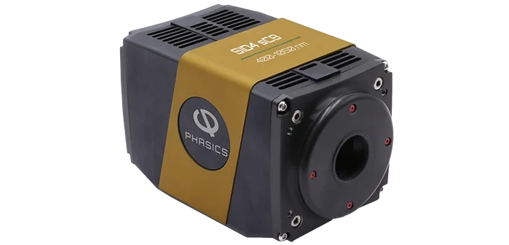
The SID4 sC8 High Resolution sCMOS Quantitative Phase Imaging Camera is engineered to deliver advanced phase measurement capabilities for both life science and material inspection applications. Leveraging cutting-edge sCMOS sensor technology, this camera provides fast, accurate, and truly quantitative phase imaging in a compact, plug-and-play format. Its design ensures seamless integration with a wide range of microscopes, making it an ideal solution for researchers and professionals seeking high sensitivity and precision in their imaging workflows.
In the realm of life sciences, the SID4 sC8 empowers biologists with label-free cell imaging, enabling the observation of live cells without the need for dyes or markers. The camera’s high sensitivity and automatic segmentation features streamline the analysis of cellular structures and dynamics, supporting advanced research in cell biology, pathology, and developmental studies. Its compatibility with fluorescence imaging further expands its versatility, allowing simultaneous phase and intensity measurements for comprehensive sample characterization.
For material scientists, the SID4 sC8 offers robust capabilities for accurate refractive index measurement, laser damage analysis, and surface characterization. The camera’s high phase spatial resolution and sub-nanometer phase accuracy facilitate detailed inspection of materials at the microscopic level. Its broad wavelength range (400-1050 nm) and compatibility with any illumination source or objective lens make it suitable for a variety of inspection tasks, from semiconductor analysis to thermal imaging.
The SID4 sC8 stands out with its single-shot phase and intensity measurement, high frame rate (up to 40 fps), and real-time processing capabilities. Its user-friendly USB 3.0 connection and compact dimensions ensure easy setup and integration into existing laboratory environments. Additionally, the camera is compatible with popular acquisition software platforms such as Metamorph, Micromanager, and NIS-Elements, supporting magnifications from x2.5 to x150. This versatility, combined with its robust performance, makes the SID4 sC8 a powerful tool for researchers and engineers demanding high-quality quantitative phase imaging.
SID4 sC8 High Resolution sCMOS Quantitative Phase Imaging Camera
Specifications
| Wavelength Range: | 400 – 1050 nm |
|---|---|
| Aperture Size (horizontal): | 16.64 mm |
| Aperture Size (vertical): | 14.04 mm |
| Phase Spatial Resolution: | 19.5 um |
| Phase & Intensity Sampling (horizontal): | 852 pixels |
| Phase & Intensity Sampling (vertical): | 720 pixels |
| Phase Resolution (RMS): | 2 nm |
| Frame Rate: | 40 fps |
| Real-Time Processing Frequency (full Res.): | 10 Hz |
| Sensor Technology: | sCMOS |
| Connection: | USB 3.0 |
| Dimensions: | 85 x 90 x 145 mm |
| Weight: | ~1100 g |
| Magnification: | x2.5 to x150 |
Got questions about specs? Use the inquiry form to ask.
 Download Data Sheet

Features
- High-Resolution sCMOS Sensor: Delivers exceptional image quality with a large 16.64 x 14.04 mm² aperture and 852 x 720 phase & intensity sampling.
- Wide Wavelength Range: Operates across 400–1050 nm, making it suitable for diverse applications in life sciences and material inspection.
- Quantitative Phase Imaging: Provides fast, accurate, and truly quantitative phase measurements for advanced research and analysis.
- Label-Free Cell Imaging: Enables high-sensitivity imaging without the need for fluorescent or chemical labels, ideal for live cell studies.
- Automatic Segmentation: Integrated software tools for easy and precise segmentation of biological samples.
- Accurate Refractive Index Measurement: Essential for material scientists conducting surface characterization and laser damage analysis.
- Single-Shot Phase & Intensity Measurement: Captures both phase and intensity data in a single acquisition for real-time analysis.
- High Phase Spatial Resolution: Achieves 19.5 µm spatial resolution and <2 nm RMS phase resolution for detailed imaging.
- Fast Frame Rate: Records at up to 40 frames per second, with real-time processing frequency up to 10 Hz at full resolution.
- Plug & Play Compact Design: Easy integration with existing microscopes; compact (85 x 90 x 145 mm) and lightweight (~1100 g).
- USB 3.0 Connectivity: Ensures fast data transfer and simple setup with modern computers.
- Broad Compatibility: Works with any illumination source or microscope objective, and supports popular acquisition software (Metamorph, Micromanager, NIS-Elements).
- Fluorescence Imaging Compatible: Seamlessly integrates with fluorescence imaging workflows for multi-modal studies.
- Wide Magnification Range: Supports objectives from x2.5 to x150 for versatile imaging needs.
Applications
- Label-Free Live Cell Imaging
Achieve high-contrast, quantitative phase imaging of live cells without the need for fluorescent or chemical labels, preserving cell viability and enabling long-term studies. - Cell Morphology and Growth Analysis
Monitor and quantify changes in cell shape, size, and confluence for applications in cell biology, cancer research, and drug screening. - Automatic Cell Segmentation and Counting
Utilize advanced phase imaging for automated segmentation and accurate counting of cells in heterogeneous populations. - Refractive Index Mapping
Precisely measure and map the refractive index of biological samples and materials, supporting research in cytology, histology, and material science. - Laser Damage and Defect Analysis
Detect and characterize laser-induced damage, micro-cracks, and defects in optical materials and coatings with high sensitivity. - Surface Topography and Roughness Measurement
Quantitatively analyze surface features, roughness, and step heights in semiconductor wafers, MEMS, and thin films. - Fluorescence and Phase Imaging Integration
Combine quantitative phase imaging with fluorescence microscopy for comprehensive multi-modal analysis of biological and material samples. - Dynamic Process Monitoring
Capture real-time changes in living cells, tissues, or materials during chemical reactions, thermal processes, or mechanical stress. - Quantitative Tissue Imaging
Perform label-free imaging of tissue sections for pathology, neuroscience, and developmental biology, enabling objective and reproducible analysis. - Thermal Imaging and Heat-Induced Phase Changes
Visualize and quantify phase shifts caused by temperature variations in materials and biological samples. - Optical Metrology and Quality Control
Implement high-resolution, non-contact inspection of optical components, microstructures, and thin films in research and industrial settings. - Microfluidics and Particle Tracking
Track and analyze particles, cells, or droplets in microfluidic devices with high temporal and spatial resolution. - Correlative Imaging with Any Illumination or Objective
Compatible with a wide range of microscope objectives and illumination sources, facilitating integration into existing imaging workflows. - High-Throughput Screening
Enable rapid, automated imaging for large-scale screening applications in drug discovery, toxicology, and materials research.
Frequently Asked Questions
What is the SID4 sC8 Quantitative Phase Imaging Camera?
What are the main applications of the SID4 sC8 camera?
Is the SID4 sC8 compatible with fluorescence imaging and various microscope objectives?
What are the key technical specifications of the SID4 sC8 camera?
What software is the SID4 sC8 compatible with?
How does the SID4 sC8 enable label-free cell imaging?
What are the advantages of using the SID4 sC8 over traditional imaging cameras?
Got more questions? Use the inquiry form to ask the supplier directly.
Your inquiry has been received.
Create an account by adding a password
Why create an account?
- Auto-complete inquiry forms
- View and manage all your past messages
- Save products to your favorites
- Close your account anytime — no hassle