
Description
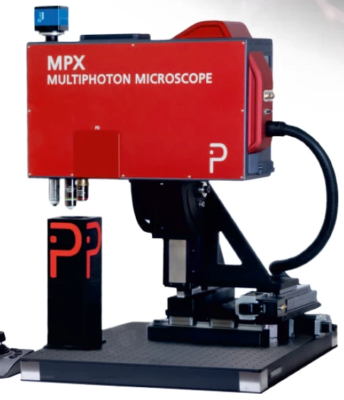
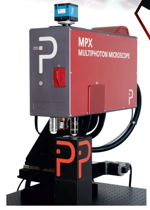
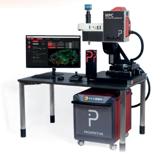
MPX-Series Multiphoton Microscopes offer a revolutionary approach to imaging with their turn-key, versatile, and multimodal capabilities. Designed for comprehensive 360° imaging, these microscopes are highly modular, allowing for extensive customization with various models, options, and accessories. Whether you are working with zebrafish eyes, drosophila brains, or mouse heads, the MPX-series provides unparalleled imaging solutions for a wide range of biological specimens.
The MPX-series is engineered to support both in-vitro and in-vivo imaging, making it an ideal choice for researchers in fields such as pathology, cancer research, and neuroscience. With its ability to perform label-free imaging and IHC/ICC, the MPX-series ensures non-destructive, deep tissue imaging. This makes it particularly suitable for applications involving spheroids, organoids, and neuronal networks, providing detailed insights into complex biological systems.
Equipped with tunable wavelengths ranging from 765 nm to 940 nm and a fixed wavelength of 1030 nm, the MPX-series offers flexibility and precision in imaging. The built-in femtosecond laser or 3-photon-ready configuration ensures high-quality imaging with minimal dark count rates. The flexible 360° scanhead accommodates inverted, upright, or oblique angle imaging, enhancing the versatility of the system for various experimental setups.
Designed with user-friendliness in mind, the MPX-series is compact and air-cooled, making it a practical addition to any laboratory. The integrated workspace illumination and motorized modality change further streamline the imaging process, allowing researchers to focus on their experiments without unnecessary interruptions. With high NA 2" detection optics and ultra-sensitive GAASP PMTs or SiPMs, the MPX-series ensures exceptional image quality and sensitivity.
In summary, the MPX-series Multiphoton Microscopes are a powerful tool for researchers seeking advanced imaging capabilities. Their modular design, combined with cutting-edge technology, provides a comprehensive solution for a wide array of imaging needs, from basic research to complex tissue engineering and bioprinting applications.
MPX-Series Multiphoton Microscopes: Versatile 360° Imaging for Research
Specifications
| Laser Source Wavelength: | 1030 nm |
|---|---|
| Laser Repetition Rate: | 100 MHz |
| Laser Average Power: | 600 mW |
| Laser Pulse Duration (on Sample): | 140 fs |
| Laser Power Control Range: | 0.5 – 100 % |
| Lateral Scan Type: | Galvo, Resonant |
| Z Scan Type: | Not Specified |
| Scan Size: | 2048 – 2048 pixels |
| Objective Revolver: | Yes |
| Signal Detection: | PMT(s), HDD(s), SiPM(s), SPAD(s), sCMOS |
| Pixel Dwell Time: | 44 ns |
| Fluorescence Camera Dark Current: | 15 e- / pixel / s |
| Fluorescence Camera Spectral Range: | 370 nm - 1100 nm |
| Fluorescence Camera Quantum Efficiency: | up to 80 % |
| Fluorescence Camera Readout Noise: | 2.1 med e- |
| Fluorescence Camera Pixel Size: | 6.5 µm |
| Scan Size: | Up to 2048 x 2048 pixels |
| Scan Zoom: | 1x to 99x |
| Beam Diameter: | 22 mm |
| Single Wavelength: | 780 / 920 / 1030 nm |
| Scan Speed: | 30 fps at 512 x 512 pixels (8 kHz resonant galvo CRS8K) |
| Pixel Dwell Time: | 0.8 to 32 µs |
| Scan Speed: | 0.3 fps at 2048 x 2048 pixels |
| Scan Speed: | 4.6 fps at 512 x 512 pixels |
| Laser Source: | built-in femtosecond lasers |
| Excitation Wavelengths: | 765 nm - 940 nm & 1030 nm |
| Second Wavelength: | 765 - 940 nm (tunable) |
| First Wavelength: | 1030 nm |
Got questions about specs? Use the inquiry form to ask.
 Download Data Sheet

Features
- Turn-Key and Versatile: The MPX-Series Multiphoton Microscopes are highly modular, offering customizable models with various options and accessories.
- Multimodal 360° Imaging: Provides comprehensive imaging capabilities, including widefield, multiphoton, and label-free imaging.
- Broad Range of Applications: Suitable for 2D/3D/4D imaging, in-vitro & in-vivo studies, label-free & IHC/ICC, deep tissue imaging, pathology, cancer research, neuroscience, optogenetics, whole slide imaging, spheroids/organoids, and tissue engineering & bioprinting.
- Tunable Wavelengths: Excitation wavelengths range from 765 nm to 940 nm and 1030 nm, with built-in femtosecond lasers or 3-photon-ready options.
- High NA Detection Optics: Features high numerical aperture 2" detection optics with up to 4 ultra-sensitive GaAsP PMTs or SiPMs, offering low dark count rates in epi or transmission modes.
- Flexible Scanhead: Allows for inverted, upright, or oblique angle imaging with a 360° scanhead.
- Integrated Workspace Illumination: Includes motorized modality change and a motorized 3-position objective revolver for enhanced user convenience.
- User-Friendly and Compact: Designed to be compact, air-cooled, and easy to use, making it ideal for various laboratory settings.
- Advanced Specifications:
- Single wavelength options: 780 / 920 / 1030 nm
- Laser power control with scan speeds of up to 30 fps at 512 x 512 pixels
- High-efficiency collection optics for fluorescence photon detection
- Widefield fluorescence imaging with white-light LED or up to 9 channel solid-state illumination
- sCMOS monochrome fluorescence camera with high quantum efficiency and low readout noise
Applications
Applications for MPX-Series Multiphoton Microscopes
- Multiphoton fluorescence microscopy for high-resolution 2D, 3D, and 4D imaging of living cells, tissues, organoids, and intact model organisms.
- Deep tissue imaging of thick biological specimens, including brain tissue, kidney, mouse head sections, zebrafish, drosophila brain, and cleared or uncleared samples.
- In vivo imaging for non-destructive observation of living organisms, including zebrafish eye imaging, zebrafish development studies, intravital microscopy, and live animal physiology research.
- In vitro cell imaging for label-free and fluorescence-based analysis of cultured cells, co-cultures, primary cells, stem cells, and engineered cellular models.
- 3D spheroid and organoid imaging for cancer biology, drug discovery, developmental biology, regenerative medicine, and disease modeling.
- Neuroscience imaging for neuronal network visualization, brain slice imaging, calcium imaging, synaptic activity studies, dendritic spine analysis, neurodevelopment research, and neurodegeneration studies.
- Optogenetics applications for light-based stimulation and functional interrogation of neuronal circuits, cells, and tissue models when configured with optogenetics options.
- Whole slide imaging of large biological specimens and tissue sections, including mouse head, kidney, zebrafish, brain tissue, pathology samples, and histology slides.
- Pathology and cancer research for tumor microenvironment imaging, tumor spheroids, cancer organoids, biopsy analysis, metastasis studies, and label-free tissue morphology assessment.
- IHC and ICC fluorescence imaging for immunohistochemistry and immunocytochemistry workflows using fluorescent markers, antibody staining, and multiplexed labeling.
- Label-free microscopy for non-destructive imaging of cells, tissues, extracellular matrix, and biomaterials without exogenous fluorescent labels.
- SHG and THG imaging for label-free visualization of collagen, extracellular matrix, muscle, connective tissue, myelin, interfaces, and structural organization in biological and engineered materials.
- CARS and SRS microscopy for chemically selective label-free imaging of lipids, proteins, cellular metabolism, myelin, drug distribution, and tissue composition when configured with nonlinear Raman imaging modules.
- FLIM imaging for fluorescence lifetime microscopy, metabolic imaging, FRET analysis, autofluorescence studies, NADH/FAD imaging, and quantitative cellular microenvironment analysis.
- Developmental biology for time-lapse imaging of embryos, zebrafish, drosophila, organoids, and morphogenesis in living biological systems.
- Regenerative medicine research for monitoring stem cell differentiation, tissue maturation, engineered constructs, wound healing models, and organoid development.
- Tissue engineering and bioprinting for imaging 3D scaffolds, hydrogels, biomaterials, printed tissues, cell infiltration, matrix remodeling, and vascularization.
- Drug discovery and toxicity screening for high-content imaging of spheroids, organoids, neuronal cultures, cancer models, and live-cell response to therapeutics.
- Live-cell imaging for longitudinal, low-phototoxicity observation of cellular dynamics, migration, proliferation, morphology, intracellular signaling, and cell-cell interactions.
- Calcium and functional imaging of neuronal networks, cardiac models, organoids, and excitable tissues using genetically encoded or chemical fluorescence indicators.
- Autofluorescence imaging of endogenous fluorophores in cells and tissues for label-free analysis of metabolism, structure, and disease-related changes.
- Large-volume volumetric imaging for 3D reconstruction of tissue architecture, organs, model organisms, thick specimens, and multi-layer biological structures.
- Intravital microscopy for studying immune cell trafficking, vascular dynamics, tumor progression, tissue remodeling, and physiological processes in living animals.
- Vascular imaging for angiogenesis studies, blood vessel morphology, perfusion analysis, tumor vascularization, zebrafish vasculature, and tissue engineering vascular networks.
- Kidney research for imaging renal tissue morphology, nephron structure, glomeruli, tubules, vascular components, and disease models.
- Brain and neural tissue imaging for mouse brain, drosophi
Frequently Asked Questions
What is the MPX-Series multiphoton microscope used for?
What is the difference between the MPX-FIXED and MPX-TUNE multiphoton microscope models?
What excitation wavelengths and laser specifications does the MPX-Series support?
Can the MPX-Series perform label-free, widefield, and multiphoton fluorescence imaging?
What detectors are available for the MPX-Series multiphoton microscope?
What scan speeds and imaging resolution does the MPX-Series provide?
How does the MPX-Series compare with other multiphoton microscopes?
Got more questions? Use the inquiry form to ask the supplier directly.
Similar Products
Your inquiry has been received.
Create an account by adding a password
Why create an account?
- Auto-complete inquiry forms
- View and manage all your past messages
- Save products to your favorites
- Close your account anytime — no hassle