
Description
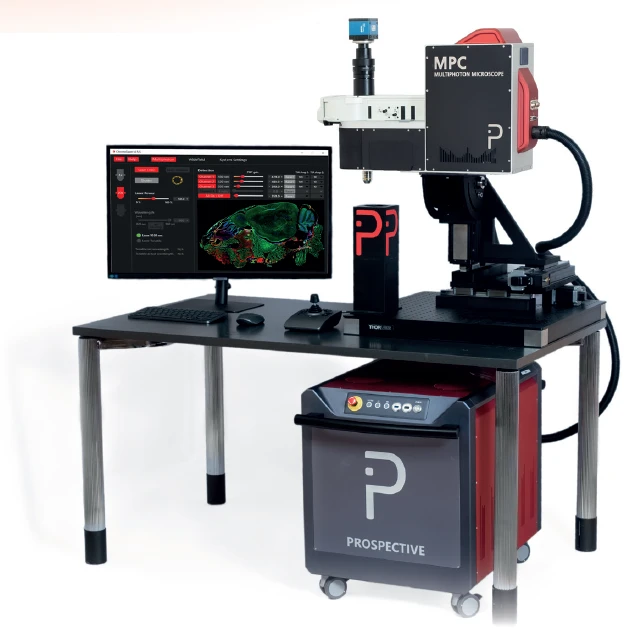
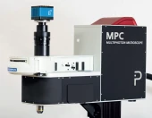
The MPC-series multiphoton multimodal imaging platform represents the pinnacle of modular imaging technology, offering unparalleled flexibility and customization. Designed to interface seamlessly with any femtosecond laser, this platform supports both 2- and 3-photon imaging, making it an ideal choice for researchers who demand precision and adaptability. Whether you choose to integrate your own laser or opt for Prospective Instruments' integrated femtosecond laser, the MPC-series ensures a tailored fit for your specific imaging needs.
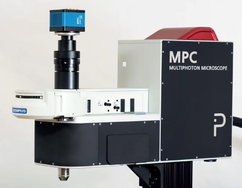
One of the standout features of the MPC-series is its highly configurable scanhead, which can be installed on an XYZ-stage in any orientation—upright, inverted, or even at oblique angles. This flexibility allows users to design a multiphoton microscope that aligns perfectly with their research objectives and budget constraints. The modular design not only facilitates easy configuration but also supports future upgrades, ensuring that your investment remains cutting-edge as new technologies emerge.
The MPC-series seamlessly combines the integrated design of the MPX-series with the versatility of fluorescence microscopes. By utilizing the Evident® (Olympus) fluorescence illuminator, users can enhance their setup with various components from the Evident® (Olympus) ecosystem, such as tube lenses or filter wheels. This integration empowers researchers to expand their imaging capabilities and adapt to evolving scientific demands.
In summary, the MPC-series multiphoton microscopes are a turn-key solution that offers unmatched flexibility and compactness. With its customizable features and compatibility with a broad range of laser sources and fluorescence detection systems, the MPC-series is poised to support a wide array of research applications, from deep tissue imaging to whole slide imaging and beyond. This platform is not just an imaging tool; it's a gateway to new scientific discoveries.
MPC-Series Multiphoton Microscope: Customizable, Multimodal Imaging Platform
Specifications
| Laser Source Wavelength: | 680 nm |
|---|---|
| Laser Power Control Range: | 0.5 – 100 % |
| Lateral Scan Type: | Galvo, Resonant |
| Z Scan Type: | Translation Stage |
| Scan Size: | 2048 – 2058 pixels |
| Objective Revolver: | Yes |
| Signal Detection: | PMT(s), HDD(s), SiPM(s), SPAD(s) |
| Wavelength Range: | 680nm to 1750nm |
| Detection: | Up to 4 PMTs, HDDs, SiPMs or SPADs |
| Field Of View (FOV): | 20 mm diagonal square (max) at the intermediate image plane |
Got questions about specs? Use the inquiry form to ask.
 Download Data Sheet

Features
- 2D/3D/4D Imaging: Advanced imaging capabilities for comprehensive analysis.
- In-vitro & In-vivo: Suitable for both laboratory and live organism studies.
- Whole Slide Imaging: Capture entire slides for detailed examination.
- Label-free & IHC/ICC: Compatible with both label-free and immunohistochemistry/immunocytochemistry techniques.
- Non-destructive: Preserve samples while obtaining high-quality images.
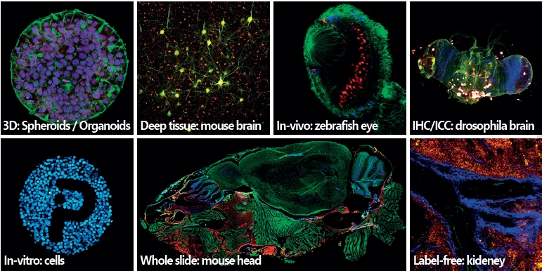
- Deep Tissue Imaging: Ideal for penetrating deeper layers of tissue.
- Pathology & Cancer: Designed for applications in pathology and cancer research.
- Neuroscience: Perfect for detailed brain studies and neurological research.
- Optogenetics: Supports optogenetic applications for advanced research.
- Tissue Engineering & Bioprinting: Facilitates studies in tissue engineering and bioprinting.
- Spheroids/Organoids: Suitable for 3D cell culture models like spheroids and organoids.
- Highly Customizable: Modular design allows for tailored configurations and future upgrades.
- 3-Photon and THG-ready: Prepared for advanced imaging techniques such as third-harmonic generation.
- Custom-designed Laser Scanning Optics: Achromatically compensated optics from 680nm to 1700nm.
- Ultra-sensitive Detection: Up to 4 GaAsP PMTs with low dark count rate for high sensitivity.
- Compatible with Evident (Olympus) Products: Integrates seamlessly with Olympus fluorescence components.
- Open Frame Design: Allows for easy upgrades and additional features.
- Optimized Fluorescence Detection: Large collection angle with 2" optics for enhanced detection.
- Broad Range of Applications: Suitable for various research fields including neuroscience, cancer, and tissue engineering.
Applications
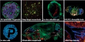
- Multiphoton microscopy for 2D, 3D, and 4D imaging: High-resolution visualization of biological samples over time using two-photon, three-photon, and advanced multiphoton imaging techniques.
- Deep tissue imaging: Non-invasive imaging of thick biological specimens, cleared tissues, organ samples, and live tissue with reduced phototoxicity and improved penetration depth.
- In-vivo imaging: Real-time imaging of living organisms and animal models, including mouse brain, zebrafish, embryos, and small model organisms.
- In-vitro imaging: High-content imaging of cultured cells, tissue sections, organotypic cultures, spheroids, organoids, and engineered tissue models.
- Whole slide imaging: Large-area scanning of histology slides, tissue sections, mouse head samples, brain slices, and pathology specimens.
- Label-free imaging: Imaging of biological structures without fluorescent labels using intrinsic contrast methods such as SHG, THG, CARS, and SRS microscopy.
- Second harmonic generation microscopy: Label-free visualization of collagen, myosin, fibrillar proteins, extracellular matrix, connective tissue, and tumor microenvironments.
- Third harmonic generation microscopy: Imaging of lipid structures, interfaces, membranes, myelin, embryos, and unstained transparent tissues.
- CARS and SRS imaging: Chemical imaging of lipids, proteins, water, drug distribution, metabolic processes, and biochemical composition in cells and tissues.
- FLIM imaging: Fluorescence lifetime imaging for metabolic studies, protein interaction analysis, FRET experiments, autofluorescence studies, and cellular microenvironment mapping.
- Pathology and cancer research: Imaging of tumor samples, cancer biopsies, tumor margins, metastasis models, extracellular matrix remodeling, angiogenesis, and cancer cell invasion.
- Digital pathology and histology: High-resolution multiphoton imaging of stained and unstained tissue sections for research pathology, tissue morphology, and diagnostic assay development.
- IHC and ICC imaging: Fluorescence imaging of immunohistochemistry and immunocytochemistry samples, including multi-marker tissue and cell analysis.
- Neuroscience imaging: Deep imaging of brain tissue, neuronal networks, dendrites, axons, synapses, glia, calcium activity, and neurovascular structures.
- Intravital brain imaging: Longitudinal imaging of neuronal activity, vascular dynamics, immune cell behavior, and disease progression in live animal models.
- Optogenetics research: Combined stimulation and imaging for studying neural circuits, functional connectivity, neuronal activation, and light-controlled biological processes.
- Zebrafish imaging: In-vivo imaging of zebrafish embryos, larvae, eye development, nervous system development, vasculature, and disease models.
- Spheroid and organoid imaging: 3D and 4D imaging of cancer spheroids, brain organoids, intestinal organoids, tumor organoids, and stem-cell-derived tissue models.
- Tissue engineering and bioprinting: Non-destructive imaging of engineered tissues, bioprinted constructs, scaffolds, hydrogels, cell-matrix interactions, and tissue maturation.
- Regenerative medicine research: Monitoring cell differentiation, tissue development, matrix formation, vascularization, and structural organization in regenerative models.
- Developmental biology: Long-term imaging of embryos, organ development, morphogenesis, cell migration, and dynamic biological processes in 3D samples.
- Stem cell research: Imaging of stem cell cultures, differentiation models, organoids, 3D matrices, and live-cell behavior in complex microenvironments.
- Cell biology and live-cell imaging: Visualization of cellular morphology, intracellular structures, signaling events, cell migration, proliferation, and cell-cell interactions.
- Vascular and angiogenesis imaging: Imaging of blood vessels, capillary networks, vascular remodeling, perfusion, endothelial behavior, and tumor angiogenesis.
- Kidney, liver, and organ imaging: Label-free and fluorescence imaging of kidney tissue, liver tissue, organ slices, disease models, and functional tissue architecture.
- Immunology and inflammation studies: Tracking immune cells, inflammatory responses, cell trafficking, tissue infiltration, and immune-tumor interactions in live or fixed samples.
- Pharmacology and drug discovery: Imaging-bas
Frequently Asked Questions
What is the MPC-Series multiphoton microscope used for?
Does the MPC-Series support 2-photon, 3-photon, and 4-photon imaging?
Can the MPC-Series multiphoton microscope perform label-free imaging?
What laser sources are compatible with the MPC-Series microscope?
How customizable is the MPC-Series multiphoton imaging platform?
What detection options are available for the MPC-Series?
Is the MPC-Series compatible with Olympus or Evident fluorescence microscope components?
Got more questions? Use the inquiry form to ask the supplier directly.
Similar Products
Your inquiry has been received.
Create an account by adding a password
Why create an account?
- Auto-complete inquiry forms
- View and manage all your past messages
- Save products to your favorites
- Close your account anytime — no hassle