Description
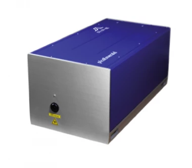
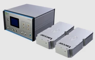
picoEmerald: Tunable Two-Color ps Light Source
The picoEmerald is a groundbreaking tunable two-color light source designed for advanced microscopy and spectroscopy applications, particularly Coherent Anti-Stokes Raman Spectroscopy (CARS) and Stimulated Raman Scattering (SRS). This innovative system integrates seamlessly into both commercial and custom-built setups, offering researchers a sophisticated tool that delivers high precision and reliability. With its capability to provide perfect temporal and spatial overlap of exit beams, the picoEmerald ensures optimal performance in imaging and spectroscopic applications.
Uniquely, the picoEmerald combines two colors in one compact, turn-key system, making it ideal for CARS and SRS applications up to video-rate speeds. The system is tunable between 700 to 1950 nm, with an energy difference ranging from 400 to 9000 cm-1. This flexibility allows researchers to tailor the light source to their specific experimental needs. The system emits 2 ps pulses with a spectral width of 10 cm-1, providing the precision required for detailed spectroscopic analysis.
Fully automated and computer-controlled, picoEmerald offers an extremely low-noise operation compared to traditional all-fiber laser systems. This makes it particularly suitable for long-term experiments where stability and consistency are crucial. The system's active and passive stabilization features further enhance its performance, ensuring that researchers can rely on it for extended periods without the need for constant adjustments.
In the realm of microscopy and spectroscopy, the picoEmerald stands out as a user-friendly, hands-off solution that simplifies complex procedures. Its integration of a laser oscillator with OPO Signal and Idler beams in a single-box design streamlines the experimental setup, allowing researchers to focus on their work rather than the intricacies of their equipment. This system is a testament to APE's commitment to advancing scientific research by providing cutting-edge tools that enhance both precision and ease of use.
APE picoEmerald Microscopy and Spectroscopy
Specifications
| Rep Rate: | Not Specified |
|---|---|
| Time Resolution: | 2000 fs |
| Laser Wavelength: | 1032 nm |
| Tunable Range: | 700 ... 1950 nm |
| Energy Difference: | 400 ... 9000 cm-1 |
| Pulse Duration: | 2 ps |
| Spectral Width: | 10 cm-1 |
| Laser Wavelength: | 1032 nm |
| Detector Area: | 10 x 10 mm |
Got questions about specs? Use the inquiry form to ask.
Features
- Two Colors in One Box: The picoEmerald system offers a two-color turn-key solution perfect for CARS and SRS applications, delivering up to video-rate speed.
- Perfect Beam Overlap: Ensures perfect temporal and spatial overlap of exit beams for optimal performance.
- Wide Tunability: Tunable between 700 - 1950 nm and 400 - 9000 cm-1 energy difference, accommodating a broad range of applications.
- Ultra-Short Pulses: Delivers 2 ps pulses with a spectral width of 10 cm-1 for precise imaging.
- Fully Automated: The system is completely automated and fully computer-controlled for ease of use.
- Low-Noise Performance: Offers extremely low-noise operation compared to all-fiber laser systems, enhancing image quality.
- Compatibility: Compatible with commercial microscopes and home-built setups, providing flexibility for various research needs.
- Stability: Actively and passively stabilized, making it ideal for long-term experiments.
- Microscopy & Spectroscopy: Designed for advanced microscopy and spectroscopy applications with a tunable two-color source.
- Enhanced Imaging Contrast: SRS microscopy with picoEmerald provides almost background-free imaging contrast and allows simple spectroscopic identification using Raman spectra databases.
- High Imaging Contrast: CARS imaging offers high contrast without labeling, sensitive to vibrational modes, and visualizes molecular vibrational contrast.
- Turn-Key System: Combines user-friendly automation features with the utility of open-architecture light sources in a single box.
- Advanced Detection Module: Includes a built-in jEOM and kSRS detection module with a large-area photodetector optimized for the VIS/NIR range.
Applications
- Coherent Anti-Stokes Raman Spectroscopy (CARS)
- Stimulated Raman Scattering Microscopy (SRS)
- Second Harmonics Imaging (SHG)
- Pump-Probe Spectroscopy
- SRS Microscopy of Vibrational Probes
- Surface Enhanced Hyper Raman Spectroscopy (SEHRS)
Frequently Asked Questions
What is the picoEmerald Microscopy and Spectroscopy system?
What are the features of the picoEmerald system?
What applications is the picoEmerald system suitable for?
What is the advantage of using the picoEmerald system for SRS microscopy?
Why is the picoEmerald system a good choice for CARS and SRS microscopy?
Got more questions? Use the RFQ form to ask the supplier directly.
Similar Products
Need pricing for this product? Send a quick inquiry
Your inquiry has been received.
Create an account by adding a password
Why create an account?
- Auto-complete inquiry forms
- View and manage all your past messages
- Save products to your favorites
- Close your account anytime — no hassle