Micro-cameras for biomedical imaging is the subject of this blog article
Introduction: Micro-Cameras for Biomedical Imaging
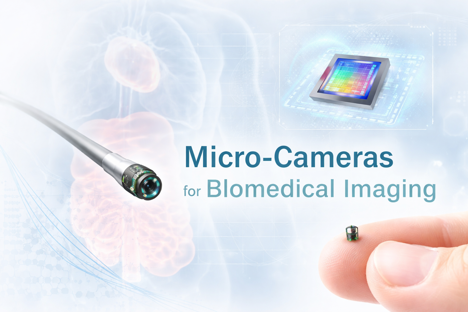
In recent years, micro-camera technology has undergone a remarkable transformation, enabling imaging systems that are smaller than a grain of rice while delivering increasingly sophisticated performance. Companies like OptaSensor GmbH are pushing the boundaries of what is possible, offering ultra-compact camera modules designed for integration into medical devices, industrial systems, and scientific instruments.
These micro-cameras are not simply scaled-down versions of conventional cameras. Instead, they represent a convergence of innovations in semiconductor imaging, micro-optics, packaging, and signal processing. The result is a new class of imaging tools that are enabling minimally invasive diagnostics, real-time monitoring, and entirely new biomedical techniques. As performance continues to improve, driven by advances in sensor sensitivity, data transmission, and system-level integration, micro-cameras are increasingly becoming foundational components in next-generation imaging platforms, expanding their role from niche diagnostic tools to ubiquitous enablers of precision medicine, smart manufacturing, and advanced scientific discovery.
The Core Technology Behind Micro-Cameras
CMOS Image Sensors: The Foundation of Miniaturization
At the heart of most modern micro-cameras lies the CMOS (Complementary Metal-Oxide-Semiconductor) image sensor. Unlike legacy CCD sensors, CMOS technology integrates photodetectors and readout electronics directly onto the same chip, significantly reducing size, power consumption, and system complexity.
This level of integration is critical for micro-cameras for biomedical imaging, where every fraction of a millimeter matters. By embedding amplification, digitization, and signal processing directly on-chip, CMOS sensors eliminate the need for bulky external electronics.
Modern micro-camera modules can be astonishingly small. For example, medical-grade devices can reach volumes below 1 mm³ while still transmitting high-quality digital signals over several meters of cable.
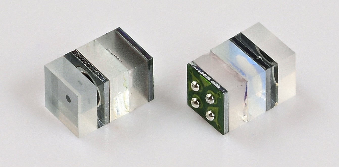
What is Inside the OS500 FOV120 Digital Micro Camera for Medical Endoscopy (Courtesy of OptaSensor GmbH)
Advanced Pixel Architectures: BSI and Beyond
To compensate for their tiny size, micro-cameras rely on advanced pixel designs such as:
- Backside Illumination (BSI) – improves light collection efficiency by illuminating the sensor from the rear side
- Microlens arrays – concentrate incoming light onto photodiodes
- Through-Silicon Via (TSV) packaging – enables vertical stacking and improved electrical routing
These technologies significantly enhance sensitivity, signal-to-noise ratio, and low-light performance, all of which are critical in biomedical environments where illumination is limited.
For instance, micro-cameras used in endoscopy often operate inside the human body, where lighting conditions are inherently challenging. High quantum efficiency and low noise are therefore essential for usable imaging.
Optics at the Millimeter Scale
Miniaturizing optics is arguably even more challenging than shrinking sensors. Micro-cameras for biomedical imaging incorporate:
- Ultra-small lenses with high numerical aperture
- Fixed-focus designs optimized for short working distances
- Wide field-of-view (FOV) configurations (often 90°–120°)
In many biomedical applications, depth of field is engineered to cover only a few millimeters to centimeters, which is sufficient for close-up imaging inside cavities or vessels.
Emerging approaches such as microlens arrays and even lensless imaging systems are further pushing the limits of compact imaging, enabling 3D reconstruction and extended depth-of-field in extremely small form factors.
Packaging and Integration: The Real Breakthrough
What truly differentiates modern micro-cameras is not just the sensor or optics, but how everything is packaged together into a highly integrated, robust, and application-ready module. In many ways, packaging has become the decisive innovation layer, transforming delicate semiconductor components into reliable tools that can operate inside the human body, industrial machinery, or harsh environments.
State-of-the-art modules integrate:
- Sensor + optics + protective window in a single unit
- Flexible or ultra-thin cabling for signal transmission
- Minimal electrical interfaces (often just power + serial data)
Beyond simple integration, advanced packaging techniques such as wafer-level optics (WLO), chip-on-tip assembly, and hermetic sealing are enabling unprecedented levels of miniaturization and durability. These approaches reduce alignment errors, improve optical efficiency, and ensure long-term stability under temperature variations, humidity, and mechanical stress. At the same time, improvements in micro-coaxial and flex cable design allow high-speed data transmission over long distances without compromising signal integrity, an essential requirement for real-time imaging.
This level of integration allows cameras to be seamlessly embedded into catheters, surgical tools, or even swallowable capsules, where space constraints are extreme and reliability is critical. In such environments, the camera is not a standalone component but an integral part of a larger system, often coexisting with illumination sources, sensors, and therapeutic elements.
For example, micro-camera modules as small as 1 × 1 × 1.6 mm can be integrated into medical devices with minimal footprint while maintaining real-time imaging capabilities. As packaging technologies continue to evolve, we are also seeing the emergence of fully self-contained imaging units with onboard processing, further reducing system complexity and enabling smarter, more autonomous imaging solutions across biomedical and industrial applications.
Micro-Cameras in Biomedical Applications
Minimally Invasive Endoscopy
The most established application is endoscopy, where micro-cameras are placed at the distal tip of a probe to visualize internal organs with high clarity and precision. This “chip-on-tip” architecture has fundamentally changed how clinicians capture images inside the body, replacing traditional fiber-bundle systems with fully digital imaging at the point of contact.
Modern CMOS-based endoscopes have largely replaced fiber-based imaging systems due to:
- Higher image quality
- Lower cost
- Easier integration and sterilization
Beyond these advantages, CMOS sensors enable features that were difficult or impossible with fiber optics, including higher frame rates, improved low-light sensitivity, and real-time image enhancement. This is particularly important in clinical environments where visibility can be limited and rapid decision-making is critical.
They are widely used across:
- Gastroenterology
- Urology
- Pulmonology
- Orthopedics
In practice, micro-cameras enable access to previously unreachable or difficult-to-navigate regions, supporting both diagnostics and surgical guidance. Their small size allows for thinner, more flexible instruments, reducing patient discomfort and recovery time while improving procedural safety. In advanced procedures, these cameras are often paired with integrated illumination (such as micro-LEDs) and image processing systems to deliver enhanced contrast, color fidelity, and even fluorescence-based visualization.
As the technology continues to evolve, micro-camera-enabled endoscopy is moving beyond simple visualization toward image-guided intervention, where real-time imaging is tightly coupled with surgical tools, robotics, and data analysis—ultimately enabling more precise, less invasive, and more effective treatments.
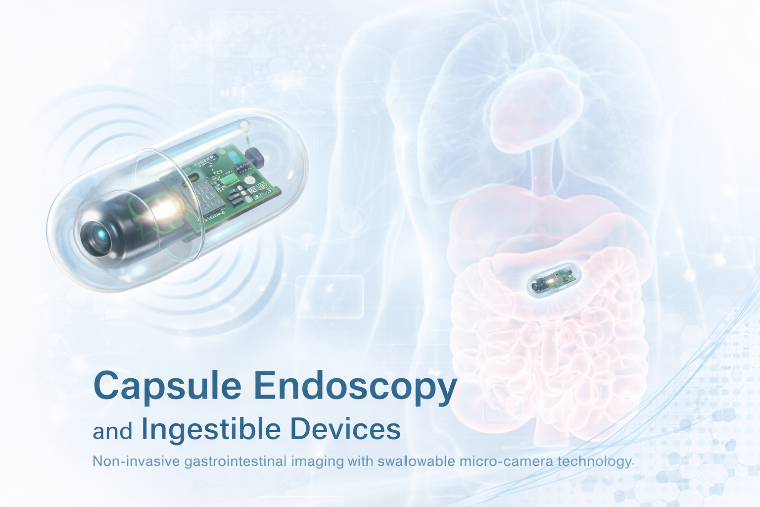
Capsule Endoscopy and Ingestible Devices
One of the most fascinating developments is the capsule endoscope – a swallowable device equipped with a micro-camera, light source, and wireless transmitter.
These systems allow non-invasive imaging of the gastrointestinal tract, eliminating the need for traditional endoscopic procedures in certain cases. Ultra-low power consumption and compact integration are key enablers of this technology.
Robotic and Image-Guided Surgery
Micro-cameras are increasingly integrated into robotic surgical systems, providing surgeons with high-resolution, real-time visual feedback.
Their small size allows:
- Multi-angle imaging
- Integration into surgical instruments
- Enhanced precision in confined spaces
This is particularly valuable in neurosurgery and microsurgery, where millimeter-scale accuracy is required.
Dental, Ophthalmic, and ENT Imaging
Micro-cameras are also widely used in:
- Dental imaging tools for intraoral inspection
- Ophthalmic devices for retinal and anterior segment imaging
- ENT applications for examining ear, nose, and throat structures
Their compact size improves patient comfort while enabling high-quality diagnostics.
Beyond Medicine: Expanding Industrial and Scientific Uses
While biomedical applications dominate, micro-cameras are rapidly expanding into other fields.
Industrial Inspection
Micro-cameras are used for:
- Inspecting turbines, engines, and pipelines
- Semiconductor wafer inspection
- Quality control in manufacturing
Their ability to access tight or hazardous spaces makes them indispensable in modern industry.
Robotics and Autonomous Systems
Compact vision modules are increasingly embedded in:
- Drones
- Micro-robots
- Autonomous inspection systems
Their low power consumption and small footprint are ideal for mobile platforms.
Scientific Instrumentation
In research environments, micro-cameras enable:
- In vivo imaging in biology
- Lab-on-chip diagnostics
- Compact microscopy systems
Emerging architectures like multi-camera arrays are even enabling gigapixel imaging by combining data from many micro-cameras.
Key Design Trade-Offs
Despite their advantages, micro-cameras involve important trade-offs:
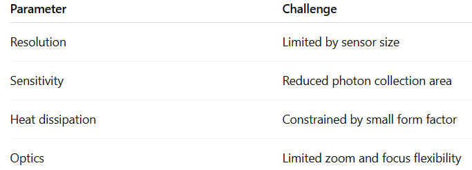
Engineers must carefully balance these factors depending on the application.
Future Trends in Micro-Camera Technology
The field continues to evolve rapidly, with several key trends emerging:
- 3D and depth imaging in ultra-compact formats
- AI-enabled edge processing within the camera module
- Improved NIR and multispectral sensitivity
- Disposable, single-use medical cameras for hygiene and cost efficiency
These trends reflect a broader shift from passive imaging toward intelligent, application-aware vision systems. Emerging depth-sensing techniques, such as time-of-flight and structured illumination, are being miniaturized to fit within millimeter-scale footprints, enabling spatial mapping in confined environments. At the same time, the integration of lightweight AI models directly on the sensor or within the camera module is allowing for real-time image enhancement, anomaly detection, and feature extraction without relying on external processing hardware.
Advances in near-infrared (NIR) and multispectral imaging are also expanding the functional capabilities of micro-cameras, particularly in biomedical applications where contrast between tissues or materials is not visible in the standard visible spectrum. This opens the door to techniques such as fluorescence-guided surgery, oxygenation monitoring, and early disease detection.
Meanwhile, the push toward disposable, single-use cameras is addressing growing concerns around sterilization, cross-contamination, and cost efficiency in healthcare environments. These systems are designed to be both high-performance and economically viable at scale, benefiting from semiconductor-style manufacturing processes.
As semiconductor and photonics technologies continue to advance, particularly in areas such as sensor fabrication, wafer-level optics, and heterogeneous integration, micro-cameras will become even more capable, compact, and intelligent. The result will be a new generation of imaging systems that not only capture data, but actively interpret and act on it, opening the door to applications that are currently unimaginable.
Conclusion
Micro-camera technology represents a powerful intersection of photonics, electronics, and precision engineering. What began as a tool for minimally invasive diagnostics has evolved into a foundational technology across medicine, industry, and scientific research.
With continued innovation from companies like OptaSensor GmbH, these tiny imaging systems are poised to play an increasingly central role in shaping the future of how we see, and understand, the world at the smallest scales.