IMA™ - Hyperspectral Fluorescence Microscope - VISNIR
Description
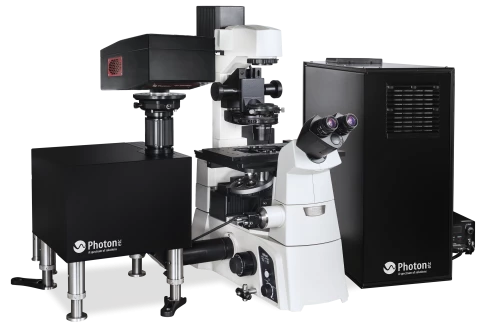
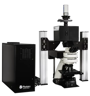
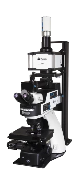
IMA™ is an ultrafast and all-in-one customizable hyperspectral microscopy platform of high spatial and spectral resolution. The completely integrated system rapidly maps diffuse reflectance, transmittance, photoluminescence, electroluminescence and fluorescence in the VIS-NIR-SWIR spectral range. Based on high throughput global imaging filters, IMA™ is faster and more efficient than scanning spectrograph-based hyperspectral systems.
Applications examples
Material science
IMA Hyperspectral Fluorescence Microscope enables complex material analysis by providing spectrally and spatially luminescence maps. Those maps can be use to study the composition, stress and inhomogeneities in a given sample. IMA can help monitor spectral information, changes in intensity of single emitters, wavelength shifts or spectral bandwidth variations. Imaging from 400 to 1700 nm, Photon etc.’s IMA™ is capable of measuring optoelectrical properties such as Open Circuit Voltage (Voc) and External Quantum Efficiency (EQE) and allows precise detection and characterization of defects in materials which is ideal for the quality control of semiconductor devices.
Life science
The spectral range covered by this Hyperspectral Fluorescence Microscope is ideal for the spatial and spectral identification and measurement of fluorophores that emit in the second biological window. With the possible integration of a dark field illumination module, it becomes an exceptional tool to detect the composition and the location of nanomaterials embedded in cells or the complex analysis of live, in vitro and unstained biological samples; the properties of organic and inorganic substances. For example, single wall nanotubes (SWNTs) emission bands are narrow (~ 20 nm) and each band corresponds to unique species (chiralities). With IMA™, it is possible to separate these species with single SWNT spatial resolution on surfaces or in live cells. This system provides attenuated tissue absorbance, higher depth of penetration and limited autofluorescence and is ideal for non destructive analysis.
IMA™ - Hyperspectral Fluorescence Microscope - VISNIR
Specifications |
|
|---|---|
| Spectral Range: | 400-1700nm |
| Spectral Resolution: | <2.5 or <4 nm |
| Detection Spectral Range: | 400-1650nm |
| Excitation Laser Wavelength: | 532nm, Other |
| Magnification: | 20x, 50x, 60x, 100x |
| Sample Stage (manual Or Motorized): | X, Y, Z |
| Microscope: | Upright or inverted |
| Spatial Resolution: | sub-micron |
| Maximum Scanning Speed: | 150 ms |
| Wavelength Absolute Accuracy: | 0.25 nm |
| Epifluorescence Filter: | Triple Filter Fluo available |
| Camera: | InGaAs, CCD, EMCCD |
| Other Filters: | Filter wheel (up to six filters) |
| Modules: | Electroluminescence, Darkfield (Oil or Dry) |
Features
IMA™ - Hyperspectral Fluorescence Microscope comes with the following important features:
- Fast global mapping (non-scanning)
- High spatial and spectral resolution
- Access to the second biological window
- Attenuated tissue absorbance
- High depth of penetration
- Low scattering
- Limited autofluorescence
- Non-destructive analysis
- Available measurements: PL, EL, reflectance, transmittance
- Customization available
- Complete system (source, microscope, camera, filter, software)
Applications
IMA™ - Hyperspectral Fluorescence Microscope is ideal for the following applications
- Characterization of solar cells
- Quality control of semiconductor devices
- Map of: composition, defects, stress, constraint, etc.
- Monitor spectral information
- Changes in intensity of single emitters
- Shifts in wavelength
- Spectral bandwidth variations
AS WELL AS IN VIVO APPLICATIONS:
- Imaging of multiplexed emitters
- Long-term sensing
An example:
Single wall nanotubes (SWNTs) emission bands are narrow (~ 20 nm) and each band corresponds to unique (n, m) species (chiralities). With IR hyperspectral microscopy, it is possible to separate these species, with single SWNT spatial resolution on surfaces, in live cells (in vivo), and in vitro.
For pricing, technical or any other questions please contact the supplier
- No registration required
- No markups, no fees
- Direct contact with supplier
-
Ships from:
Canada
-
Sold by:
-
On FindLight:
since 2015
Frequently Asked Questions
IMA™ comes with fast global mapping (non-scanning), high spatial and spectral resolution, access to the second biological window, attenuated tissue absorbance, high depth of penetration, low scattering, limited autofluorescence, non-destructive analysis, and available measurements: PL, EL, reflectance, transmittance. Customization is also available.
The spectral range covered by IMA™ is ideal for the spatial and spectral identification and measurement of fluorophores that emit in the second biological window. It can be used for the complex analysis of live, in vitro and unstained biological samples; the properties of organic and inorganic substances.
IMA™ is ideal for the characterization of solar cells, quality control of semiconductor devices, map of composition, defects, stress, constraint, etc. It can also be used for imaging of multiplexed emitters and long-term sensing.
IMA™ is an ultrafast and all-in-one customizable hyperspectral microscopy platform of high spatial and spectral resolution.
The spectral range covered by IMA™ is from 400 to 1700 nm.